The Fifth Universal Definition of Myocardial Infarction represents a major conceptual update in the way myocardial infarction is classified and diagnosed.
THE STUDY
Mills N et al. Fifth Universal Definition of Myocardial Infarction (2026) European Heart Journal, ehag101, https://doi.org/10.1093/eurheartj/ehag101 28 August 2026
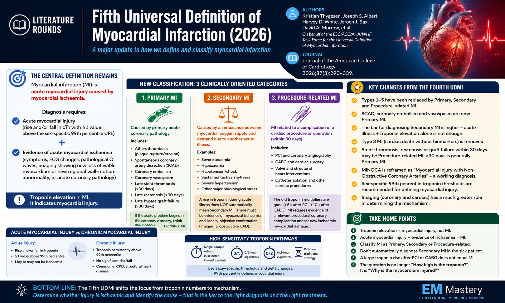
The old Type 1 - 5 classification has been replaced by a simpler and more clinically intuitive framework:
-
Primary myocardial infarction
-
Secondary myocardial infarction
-
Procedure-related myocardial infarction
An elevated troponin tells you that myocardial injury has occurred. It does not, by itself, diagnose myocardial infarction.
The clinical task is to determine whether the injury is acute, ischaemic, and if an infarction, what caused it.
The central definition remains unchanged
Myocardial infarction continues to require:
Acute myocardial injury defined by a rise and/or fall in cardiac troponin with at least one value above the sex-specific 99th percentile upper reference limit.
PLUS
Evidence of acute myocardial ischaemia. This may be demonstrated by:
-
symptoms consistent with myocardial ischaemia
-
new ischaemic ECG changes
-
development of pathological Q waves
-
imaging showing new loss of viable myocardium or a new regional wall-motion abnormality in an ischaemic pattern
-
demonstration of an acute coronary abnormality.
The presence of acute myocardial injury alone is insufficient to diagnose MI. There must also be evidence that the myocardial injury is caused by ischaemia.
A raised troponin means myocardial injury. When associated with a rise or fall it means acute myocardial injury.
Only when there is evidence that the injury is caused by myocardial ischaemia should the diagnosis of myocardial infarction be made.
Why this is important in Emergency Medicine
Troponin elevation is common in acutely unwell patients.
It may occur with:
The presence of elevated troponin in any of these settings does not automatically indicate MI.
What is causing the troponin elevation?
This is particularly relevant in patients who are physiologically stressed, where there has traditionally been a tendency to label a troponin rise as “Type 2 MI” without convincing evidence of myocardial ischaemia.
The Fifth UDMI instead categorises myocardial infarction according to its underlying mechanism.
Primary myocardial infarction
Primary MI occurs when myocardial infarction is caused by a primary acute coronary abnormality.
This includes the classical atherothrombotic event associated with plaque rupture or erosion, but the category is now broader.
Primary MI can include:
-
atherosclerotic plaque disruption and coronary thrombosis
-
spontaneous coronary artery dissection
-
coronary embolism
-
coronary vasospasm
-
late stent thrombosis
-
late restenosis
-
late bypass graft failure.
The key conceptual point is that the pathology begins within the coronary circulation itself.
Under earlier definitions, conditions such as coronary embolism, vasospasm and SCAD could sometimes be grouped under Type 2 MI because plaque rupture was absent.
Secondary myocardial infarction
Secondary MI occurs when another acute illness produces a significant imbalance between myocardial oxygen supply and demand, resulting in myocardial ischaemia and infarction.
Potential precipitants include:
The important sequence is:
Acute illness → supply-demand mismatch → myocardial ischaemia → myocardial infarction.
Compared with Primary MI:
Acute coronary pathology → myocardial ischaemia → myocardial infarction.
Secondary MI
In the previous framework, a patient with sepsis, AF with rapid ventricular response, severe anaemia or shock who developed an elevated troponin could be labelled as having Type 2 MI.
Now the patient must have evidence that the myocardial injury is ischaemic. Therefore a patient may have sepsis + troponin elevation + dynamic troponin change and have acute myocardial injury rather than myocardial infarction.
Therefore acute systemic illness can produce myocardial injury through multiple non-ischaemic mechanisms.
Supply-demand stress plus elevated troponin does not equal Secondary MI.
Imaging and assessment of underlying coronary disease assume greater importance in this setting.
A practical example
Consider a septic patient with a troponin rise from 40 to 180 ng/L.
This confirms acute myocardial injury.
If the patient has no convincing ischaemic symptoms, no dynamic ischaemic ECG changes and no new ischaemic wall-motion abnormality, the most appropriate diagnosis may be:
Acute myocardial injury associated with sepsis.
Now consider a patient with a massive gastrointestinal haemorrhage, haemoglobin of 55 g/L, chest pain, dynamic ST depression and a substantial troponin rise.
If there is evidence of significant coronary artery disease or new myocardial ischaemia, this is much more consistent with:
Secondary myocardial infarction precipitated by severe anaemia.
An acute illness does not exclude Primary MI
A patient with sepsis, major haemorrhage or another acute illness can still develop plaque rupture and atherothrombotic MI.
The presence of an obvious physiological stressor should therefore not automatically lead to a diagnosis of Secondary MI.
For example:
Sepsis + chest pain + dynamic ST changes + marked troponin rise could represent:
Persistent or recurrent myocardial ischaemia, significant ECG changes or a clinical picture suspicious for ACS should prompt consideration of coronary imaging.
Sex-specific troponin thresholds
The Fifth UDMI strengthens the recommendation to use sex-specific 99th percentile upper reference limits when defining myocardial injury. Women generally have lower 99th percentile troponin concentrations than men. Using a single threshold may therefore under-recognise myocardial injury in women.
The practical implication is that laboratories and clinicians should be aware of whether the assay in use reports sex-specific reference limits.
Acute versus chronic myocardial injury
The Fifth UDMI retains an important distinction between acute and chronic myocardial injury.
If cardiac troponin is above the 99th percentile and demonstrates a rise or fall, this is:
Acute myocardial injury.
If cardiac troponin is persistently elevated without a significant dynamic change, this is:
Chronic myocardial injury.
Chronic elevation may be seen in conditions such as:
-
chronic kidney disease
-
structural heart disease
-
chronic heart failure.
These patients may have significant cardiovascular risk, but they should not be diagnosed with acute MI simply because their troponin is elevated.
A useful diagnostic sequence is therefore:
Is troponin elevated?
If yes → myocardial injury.
Is there a rise or fall?
If yes → acute myocardial injury.
If no → chronic myocardial injury.
Is there evidence the acute injury is ischaemic?
If yes → myocardial infarction.
If no → acute non-ischaemic myocardial injury.
Procedure-related myocardial infarction
Another major change is the consolidation of the old procedural MI categories.
Previously:
-
Type 4a referred to PCI-related MI
-
Type 4b to stent thrombosis
-
Type 4c to restenosis
-
Type 5 to CABG-related MI.
These are now grouped under:
Procedure-related myocardial infarction.
This encompasses MI arising as a complication of a cardiac procedure, including:
The goal is to use a more consistent framework across both percutaneous and surgical interventions.
The 30-day procedural window
The Fifth UDMI also introduces a practical 30-day framework.
Coronary complications occurring within 30 days of an intervention may be classified as procedure-related MI when they are related to the treated coronary territory.
This may include:
Events occurring beyond 30 days are generally treated as new coronary events and are more likely to be classified as Primary MI.
The 30-day period is pragmatic rather than biological, but it aligns with traditional reporting of procedural complications.
MINOCA has been reframed
Myocardial Infarction with Non-Obstructive Coronary Arteries.
is now reframed as:
Myocardial Injury with Non-Obstructive Coronary Arteries.
Imaging becomes increasingly important
A major theme of the Fifth UDMI is that troponin should trigger diagnostic reasoning rather than terminate it.
Coronary imaging answers:
What happened in the coronary arteries?
This may involve:
Cardiac imaging answers:
What happened to the myocardium?
This may involve:
-
echocardiography
-
cardiac MRI
-
nuclear imaging.
Imaging is particularly useful when trying to differentiate:
High-sensitivity troponin pathways
The Fifth UDMI formally acknowledges contemporary high-sensitivity troponin strategies.
These include:
An important distinction is that accelerated pathways frequently use assay-specific thresholds that are not identical to the 99th percentile.
The 99th percentile defines myocardial injury.
Accelerated pathways use validated concentrations and delta changes to estimate the likelihood of acute MI.
Absolute troponin changes are generally more useful when concentrations are low or close to the 99th percentile, but any delta must be interpreted according to the specific assay.
There is no universal troponin delta that applies to every high-sensitivity assay.
What remains unchanged?
Despite the new classification, several important principles remain.
STEMI and NSTEMI are still clinically relevant.
The Primary/Secondary/Procedure-related classification describes the mechanism of infarction.
STEMI/NSTEMI describes the ECG presentation and acute management pathway.
Summary